


QIMA Life Sciences dispose de nombreux modèles in vitro ou ex vivo :
- Lignées diverses
- Cellules immuno-inflammatoires de sang périphérique humain (PBMCs et cellules purifiées : monocytes, lymphocytes, polynucléaires, etc.)
- Macrophages et cellules dendritiques différenciées de monocytes
- Kératinocytes épidermiques humains normaux (NHEK)
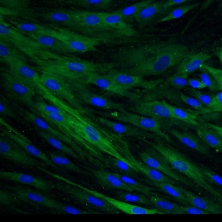
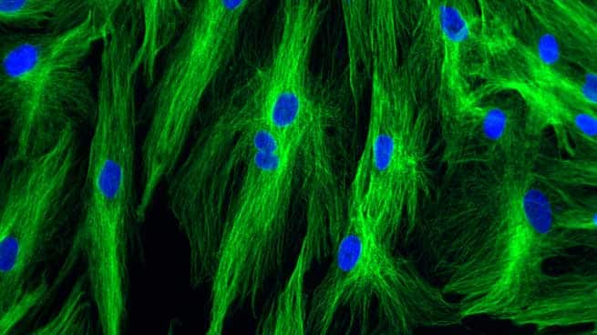
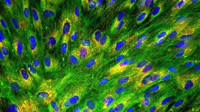
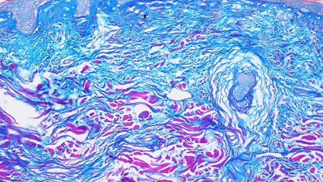
- Fibroblastes dermiques humains normaux (NHDF)
- Myofibroblastes
- Cellules souches mésenchymateuses
- Cellules endothéliales microvasculaires
- Mélanocytes épidermiques humains normaux (NHEM)
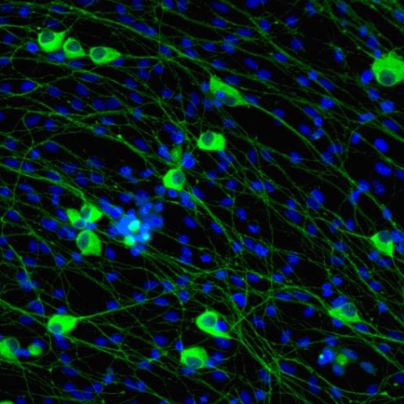
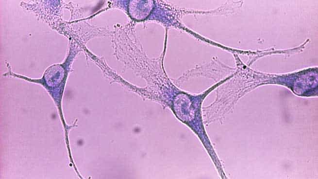
- Neurones sensitifs
- Épidermes humains recontruits (RHE)
- Peaux équivalentes
- Explants de peau (ex vivo)
sur lesquels il est possible au cas par cas d’analyser les effets procicatrisants d’actifs, formulations ou dispositifs médicaux en mesurant :
- La réponse inflammatoire, la détersion et l’immunité innée
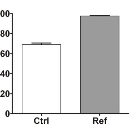
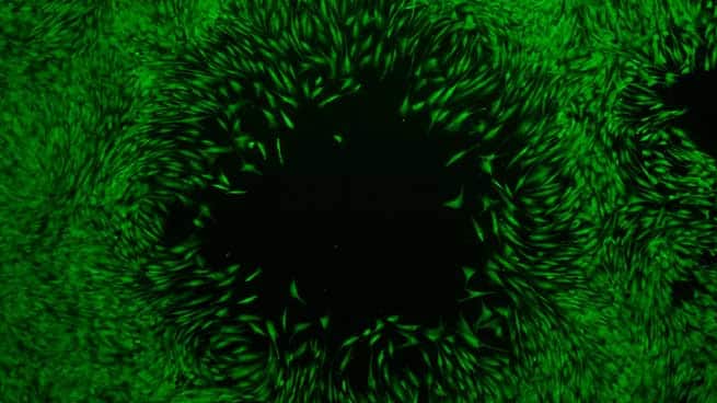
- La réepithélialisation
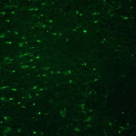
- La néovascularisation et la neuritogenèse
- La synthèse de matrice extracellulaire, la fibrose et l’interaction des myofibroblastes
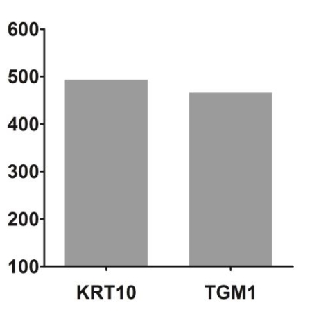
- Le remodelage dermique et la différenciation épidermique
- La pigmentation
Parmi l’ensemble des tests d’évaluation proposés par QIMA Life Sciences dans le domaine de la cicatrisation et régénération de la peau, voici quelques exemples :

Fibroblasts are potentially the key sensors of epidermis lesion through keratinocyte IL-1 signalling
Cicatrisation, DermatologieOur aim was to revisit the primary sterile epidermis lesion considering the alarmins constitutively expressed in the skin tissue and notably the IL-1 family members (IL-1fm) released by lesional keratinocytes.
At homeostasis, the non-lesional epidermis constitutes a barrier against various external aggressions…
Cicatrisation cutanée, phases de granulation et maturation
CicatrisationLa troisième phase de cicatrisation consiste à remplacer la matrice de fibrine provisoire par le tissu de granulation. Elle se déroule en plusieurs étapes: la réépithélialisation, la fibroplasie, le dépôt de collagène et l’angiogenèse.
Cicatrisation cutanée, hémostase et phase inflammatoire
CicatrisationLa phase hémostasique est la réponse immédiate à la blessure, pour stopper la perte de sang au niveau de la plaie dans les premières minutes qui suivent la lésion. Les dommages aux capillaires sanguins et l’hémorragie entraînent une vasoconstriction et la coagulation du sang.
Cicatrisation cutanée, généralités
CicatrisationLa cicatrisation cutanée est un processus naturel, dynamique et complexe impliquant de nombreux acteurs pour restaurer les structures cellulaires et tissulaires de la peau : cellules souches, cellules différenciées, follicules pileux, matrice extracellulaire (ECM), protéines, cytokines, certains microARN, ainsi que les systèmes vasculaires et nerveux et des forces mécaniques.
Study of proliferation and 3D epidermal reconstruction from foreskin, auricular and trunk keratinocytes in children
Cicatrisation, Wound healingOur studies highlight the potential of foreskin tissue for autograft applications in boys. A suitable alternative donor site for autologous cell transplantation in female paediatric burn patients remains an open question in our department. We tested the hypothesis that in vitro studies and RHE reconstructive capacities of cells from different body sites can be helpful to select an optimal site for keratinocyte isolation before considering graft protocols for girls.
Epidermal healing in burns: autologous keratinocyte transplantation as a standard procedure: update and perspective
Cicatrisation, Wound healingIn the contexte of skin graft, cell suspensions transplanted directly to the wound is an attractive process, removing the need for attachment to a membrane before transfer and avoiding one potential source of inefficiency. Choosing an optimal donor site containing cells with high proliferative capacity is essential for graft success in burns.
Foreskin-isolated keratinocytes provide successful extemporaneous autologous paediatric skin grafts
Cicatrisation, Wound healingWe report a successful method for grafting paediatric males presenting large severe burns through direct spreading of autologous foreskin keratinocytes. This alternative method is easy to implement, improves the quality of skin and minimizes associated donor site morbidity. in vitro studies have highlighted the potential of foreskin tissue for graft applications and could help in tissue selection with the prospect of grafting burns for girls.
Quantitative and qualitative study in keratinocytes from foreskin in children: Perspective application in paediatric burns
Cicatrisation, Wound healingKeratinocytes from foreskin have a high capacity for division. A potential source of cells to provide coverage in paediatric burns.
Cultured keratinocyte cells from foreskin and future application for burns in children
Cicatrisation, Wound healingThe keratinocytes resulting from foreskin have a high capacity of division. These cells can divide a long time before differentiation. The observations enable us to propose with our patients the keratinocytes from foreskin for wound healing especially for burns in children.
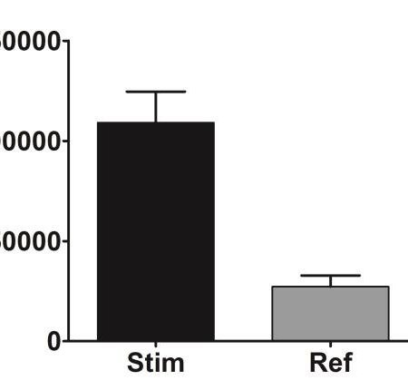
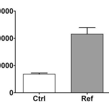
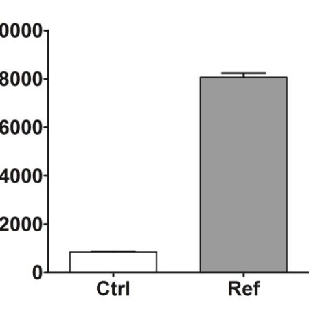
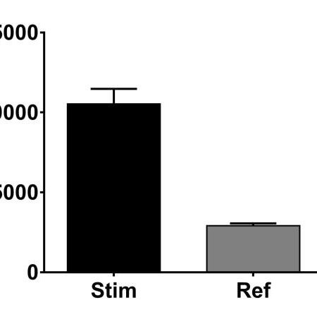
Stimulation of the proliferation of human dermal fibroblasts in vitro by a lipidocolloid dressing
Cicatrisation, Wound healingThe effect of Urgotul on normal human dermal fibroblast proliferation was studied in vitro and compared with that of two other dressing: Mepitel and Tulle Gras.