


WHAT YOU WILL LEARN IN THIS ARTICLE:
- What Are In Vitro and Ex vivo Models?
- Alternative Models for Animal Experimentation
- In Vitro vs. Ex Vivo: Differences and Similarities
- Is One of The Two Models Better Than The Other?
Scientists are continuously trying to develop better models and tools to study the biology of the skin so that we can better predict the efficacy and safety not only of cosmetic and dermatological products but also of pharmaceuticals. Among these are the 3D in vitro models, such as the Reconstructed Human Epidermis, which have significant advantages over traditional 2D in vitro models.
However, in recent years, ex vivo skin models have gained much popularity since they are very frequently regarded as “the most representative model of the human skin”.
In this article, we will delve into the similarities and differences between in vitro and ex vivo models of skin, and discuss their suitability for different research scenarios.
What Are In Vitro and Ex Vivo Models?
Before we explore the comparison between these models, let’s start with their definitions.
“In vitro” and “Ex vivo” are two Latin expressions that describe the test environment of experiments.
In vitro literally translates from Latin as “in glass.” These methods involve experimenting with cells outside a living organism. The original reference to glass is quite literal since in vitro experiments were historically conducted in Petri dishes or test tubes, made of glass. In vitro cultured cells are purified and isolated from their natural biological environment.
It is worth noting that increasingly complex in vitro models are continuously being developed. Not only they can include more than just one type of cell (in which case they will be known as co-cultures, in contrast to mono-cultures), but they can also adopt 3D configurations (such as the skin-reconstructed models or organoids).
Ex vivo literally translates from Latin as “out of the living.” In these experiments, living tissues are directly taken from a living organism and immediately studied in a laboratory setting with minimal alterations to the organism’s natural conditions. An example of this is the use of human skin explants derived from surgical procedures.
Alternative Models for Animal Experimentation
Another key aspect of in vitro and ex vivo models is that they offer a great alternative to animal testing.
This consideration is essential for cosmetic research in the EU and other countries that have fully banned animal testing in cosmetics, or for companies aiming to market their products in these regions.
In dermatology and pharmacology, while in vivo studies are still typically required before testing drugs and formulas in humans, ex vivo models have significantly contributed to generating valuable data that better predict in vivo efficacy and aid in selecting the best lead drug candidates. As a result, they help save time and money, thereby accelerating drug development.
In Vitro vs. Ex Vivo: Differences and Similarities
While both approaches occur outside living organisms, they have significant differences.
The table below compares some of the most important traits of in vitro and ex vivo models. It is essential to note that the term “in vitro models” encompasses various models, including 2D mono-cellular cultures, 2D co-culture models, and 3D organotypic models, making generalizations challenging.
You may also be interested in
Skin Barrier & Hydration

| In vitro | Ex vivo | |
|---|---|---|
| Ethical considerations | Alternative to animal testing. | Alternative to animal testing. Allows experimental interventions and analyses that would be difficult to perform on patients. |
| Spacial structure | In vitro cultures can be prepared in 2D and 3D arrangements. 3D cultures resemble skin tissues. | 3D structure similar to live skin tissues. |
| Cell populations and cell environment | Reconstructed models tipically contain only keratinocytes, melanocytes, and fibroblasts, lacking important cell populations and ECM components. Cells are isolated, separated, and purified from their normal biological surrounding. While this allows for detailed cellular and molecular analysis, it may overlook crucial intercellular interactions during the analysis. | Tissue’s resident cell populations are maintained as well as their interactions with the extracellular matrix (containing fibroblasts, collagen, elastin glycosaminoglycans, etc.). |
| Skin appendages | They miss entire skin appendages like hair follicles and sebaceous glands. | They contain hair follicles and sebaceous glands. |
| Physiology | Less physiologically similar to live skin tissues. | More physiologically similar to live skin tissues. |
| Sourcing | Isolated cells used for in vitro cultures can be amplified in culture under appropriate conditions for future reuse. | Preservation over a prolonged period is challenging, but sufficient human skin supply is possible through proper logistics and collaborations with hospitals and clinics that generate excess skin during elective plastic surgeries. |
| Production and set-up | Preparing and reconstructing in vitro models can be time-consuming. | Easy to set up and can be used immediately. |
| Inherent varibility | Defined. Cell populations are selected and manipulated in a way that enables greater reproductibility in monolayer cultures or the reconstructed models. | Undefined. It is mainly related to the intrinsical interindividual variability from which the explants are obtained. Many sample properties cannot be controlled, since they depend entirely on the biopsied tissue. For instance, characteristics that are dependent on the age and living patterns of the donors (e.g., sunlight exposure, treatment history, and allergy). |
| Biological responses to a treatment or stress | More reactive due to the simplified nature of the model. | Less reactive due to the robustness and complexity of the model. |
| Lifespan | Limited, with primary cell cultures and reconstructed models usable for a few days to a few weeks during the maturation period. | Used within a period of 10 to 14 days based on culture conditions. |
| Parameters manipulation | Maturation time can be controlled. | Maturation time is not controlled. |
| Genetic engineering | Extensive. Targeted genetic engineering is possible in isolated cell populations (gene knockout, transgene, CRISPR). This provides more powerful molecular tools for mechanistic research. | Limited. Extensive targeted genetic engineering is not yet possible. Molecular tools for mechanistic research are currently limited to gene silencing. |
| Diseased models | Mimicked in the laboratory through chemical and biological stimuli, making them available as needed. | Real diseased skin but depend on sourcing and supply availability. |
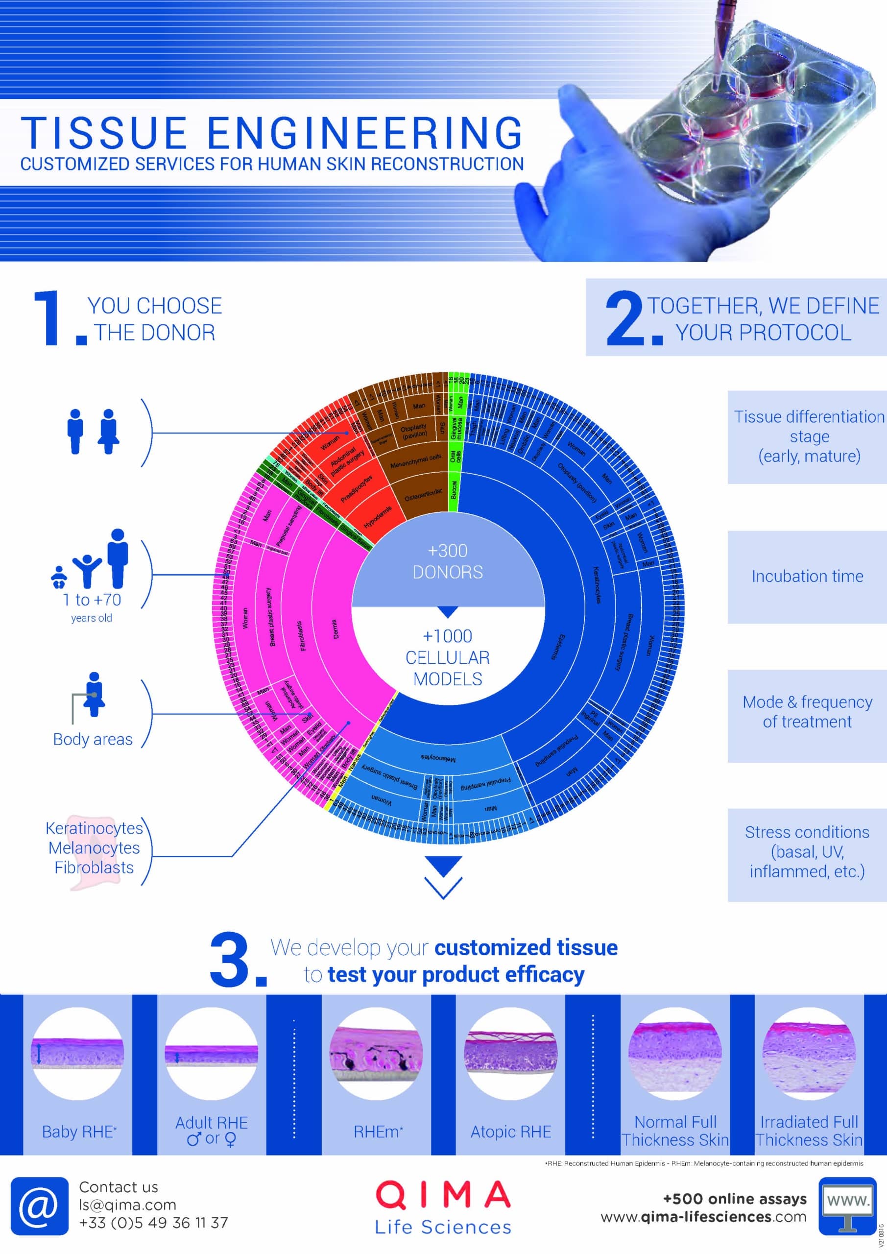
Learn more about our
Tissue Engineering services
Is One of The Two Models Better Than The Other?
The choice between in vitro and ex vivo models largely depends on the specific research goals.
Ex vivo human skin stand out as the most representative model of human skin. They are particularly valuable when studying phenomena like transdermal delivery after topical application or when investigating the effect of products on skin integrity and tissue morphology.
In contrast, in vitro models offer versatility, making them suitable for testing products on different skin types simultaneously (they enable higher throughput testing) or when a deeper understanding of the mechanism of action is required (simplified systems enable a more in-depth study of specific cell populations).
Skin explants do not fully replace in vitro models; instead, they complement each other. Combining various models provides more robust insights into skin physiology, specific skin disorders, and their potential treatments.
“Ideally, the selected models should not only be pragmatic, accessible, affordable, and instructive but should be both physiologically and clinically relevant; that is, they should faithfully reflect at least some of the complexity of the human integument under well-defined, standardized and therefore reproducible preclinical research conditions”.
(Zhou L., Zhang X., Paus R. & Zhongfa L., Differentiation, 2018.)